Advanced Imaging for Molecular Interactions and Cellular Dynamics
Functional Imaging combines advanced microscopy techniques to study molecular interactions, diffusion, and cellular dynamics with exceptional precision. Using state-of-the-art technologies such as FRET, FLIM, FRAP, and FCS, this platform enables researchers to delve into the functional and structural aspects of biological systems.

Applications
FRET (Fluorescence Resonance Energy Transfer):
FRET detects proximity and interactions between molecules at nanometer distances, providing insights into molecular interactions with unparalleled sensitivity. By measuring energy transfer between a donor and an acceptor fluorophore, FRET is ideal for studying molecular assemblies, signaling pathways, and conformational changes.
FLIM (Fluorescence Lifetime Imaging Microscopy):
FLIM measures fluorescence lifetime variations to analyze molecule proximity, pH changes, or polarity. At IBDM, FLIM systems (LSM 880 and Leica SP8) allow:
- Fast molecular interaction tracking via FLIM-FRET.
- Detection of metabolic state changes using biosensors.
- Multi-fluorophore separation using lifetime contrast.
FRAP (Fluorescence Recovery After Photobleaching):
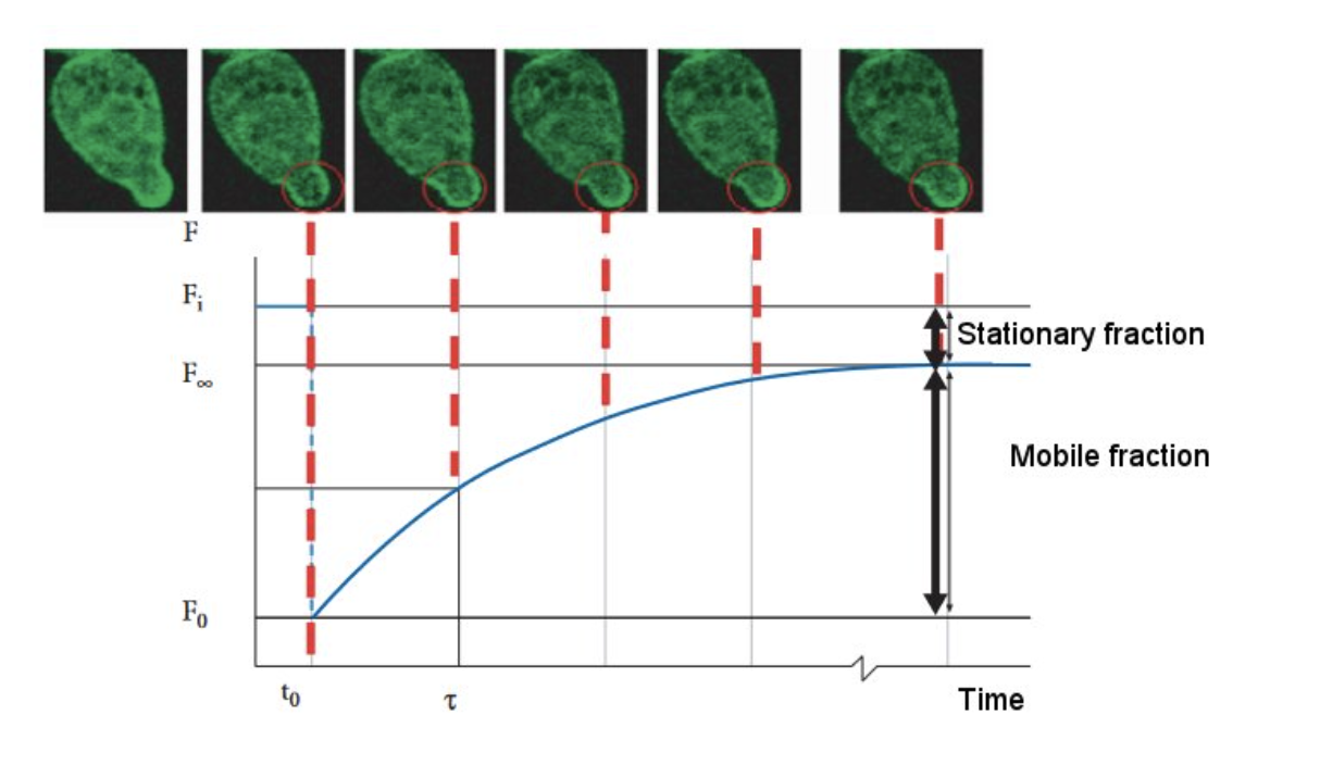
FRAP quantifies diffusion and binding dynamics of fluorescent molecules. At IBDM, FRAP acquisition on spinning disks or two-photon systems ensures precise photobleaching control for dynamic studies.
FCS (Fluorescence Correlation Spectroscopy):
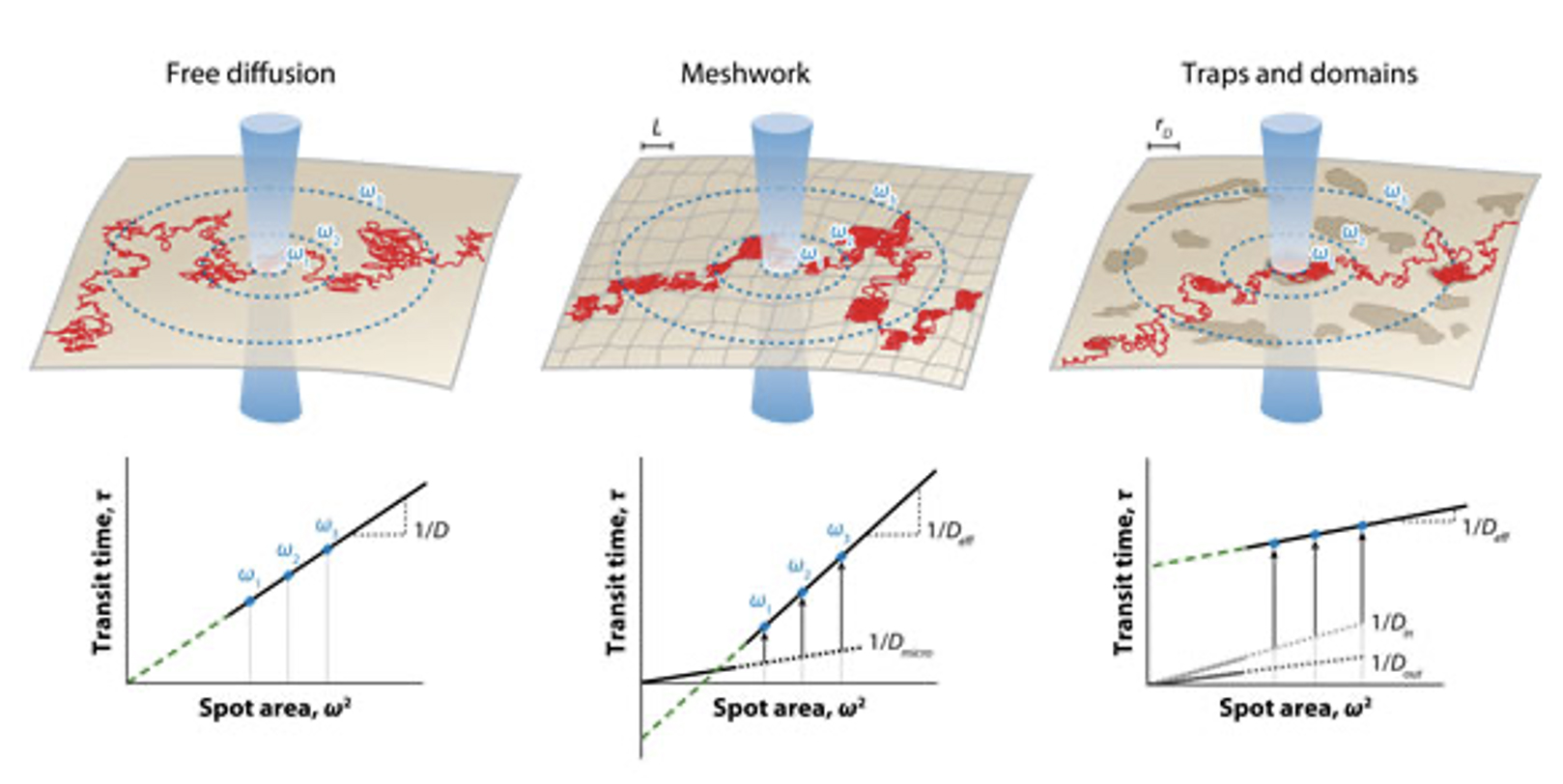
The CIML ImagImm platform has set previously an original approach based on FCS diffusion measurements to probe the lateral organization and dynamics of membranes in living cells, but performed at different spatial scales, namely the spot-variation FCS (svFCS). This setup provides FCS measurements with optimal sensitivity; it is combined with dedicated analytical tools of the recorded data. The FCS setup has been also combined with a STED illumination module.
The different svFCS modalities extended the measurements to quantify transient molecular association of cellular components with the plasma membrane of living cells (Fluorescence Correlation Spectroscopy Diffusion Laws to probe the Submicron Cell Membrane Organisation, Biophys J, 2005, 4029-4042) and (Detecting Nanodomains in Living Cell Membrane by Fluorescence Correlation Spectroscopy, Annual Review of Physical Chemistry, Volume 62, 2011).


FRET analyzes molecular proximity and interactions with nanometer sensitivity.

FLIM measures fluorescence lifetime to detect environmental and molecular changes.

FRAP quantifies diffusion and binding dynamics in live samples.

FCS studies molecular concentration, diffusion, and binding with high precision.

Combines widefield, spinning disk, and confocal microscopy for dynamic imaging.


