Linking Live-Cell Imaging to Ultrastructural Details
Correlative Light and Electron Microscopy (CLEM) combines the live-cell imaging capabilities of fluorescent light microscopy with the high-resolution structural details of electron microscopy.
This innovative approach enables precise localization of molecules within biological samples while preserving their ultrastructural context.
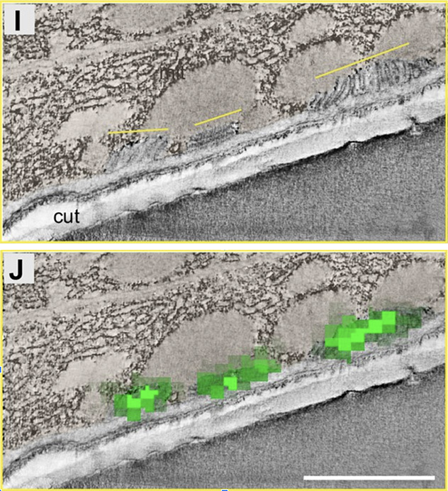
* CLEM to localize the VHA-5 protein in the Meisosomes of C. elegans (from Aggad, Brouilly et al., 2023)

Applications
Pre-embedding CLEM targets rare events by acquiring light microscopy images on samples before dehydration, resin embedding, and sectioning. This method uses the high throughput of light microscopy to screen samples at the millimeter scale, identifying regions of interest (ROI). These ROIs can then be mapped using gridded plates or natural landmarks such as nuclei or blood vessels. After electron microscopy preparation, including contrasting and resin embedding, targeted trimming and ultra-thin sectioning reveal the ultrastructure of the exact same cells. However, due to distortions during EM procedures, correlation accuracy remains at the cellular scale.
Post-embedding CLEM allows precise molecular localization at the sub-cellular scale within the ultrastructural context. Here, light microscopy acquisition is performed directly on the EM section using dedicated protocols to preserve fluorescence. Advanced protocols even enable super-resolution light microscopy, minimizing the resolution gap between light and electron microscopy techniques.

Combines live-cell imaging with high-resolution electron microscopy.

Post-embedding CLEM localizes molecules at sub-cellular scale.

Pre-embedding CLEM targets rare events and maps regions of interest.

Advanced protocols preserve fluorescence for light and super-resolution microscopy.

Links molecular localization to ultrastructural biological context effectively.


